
“My dog has a lump! Is it cancerous?” Understandably, many owners worry about their pet’s health and the possibility of cancer when they find a new skin growth or wart.
If you find a lump on your dog’s body, don’t panic right away. Many lumps and bumps are harmless or easily treated by a vet. So what types of dog lumps are there, and how can you tell them apart?
This article is heavy on veterinarian terminology.
Common Causes of Lump on a Dog’s Skin
If you find a new lump or bump on your dog’s skin, try not to panic right away – there are many reasons that your dog might have a growth! While dogs can get cancerous growths, it’s important to know that many are treatable and caused by much simpler factors.
Hematomas
A hematoma is a collection of blood outside of the blood vessels. Most commonly, a hematoma is the result of an injury to the blood vessel wall. Injuries can occur with any type of blood vessel, including arteries, veins, and small capillaries. A blood vessel injury causes blood to seep out of the vessel and into the surrounding tissues. In most cases, hematomas show up as purplish bruises under the skin. Hematomas can also form a mass or lump if enough blood collects into the area.
In dogs, hematomas are most common on the ears. If your dog has an aural hematoma, it’s usually because of head shaking or ear scratching, typically as a result of an ear infection or ear mites. As the hematoma develops, you may find that your dog’s earflap becomes heavy, swollen, and very uncomfortable. As well as this, your dog may hold their head to one side to try to relieve their discomfort. Large hematomas often require draining by a vet. Without treatment, large aural hematomas are likely to scar over or refill in the future.
Abscesses
An abscess is a pocket of pus within the tissues, typically as a result of a bacterial infection. In rare cases, some parasites cause abscesses. Signs of skin abscess include pain, warmth, redness, and swelling. Some abscesses are palpable, meaning that movement of the pus inside can be felt when you touch the abscess. The other type of abscess is an internal abscess. These are harder to diagnose and are often more serious, as they can form inside organs or in the spaces between the organs. The symptoms of an internal abscess include a high temperature, discomfort in the affected area, vomiting, loss of appetite, and weight loss.
One of the most common causes of abscesses in dogs is an animal bite. A bite injury introduces harmful bacteria into the wound. This leads to infection, and depending on the type of bacteria and the severity of the bite, an abscess can quickly develop. Other causes of abscesses in dogs include penetrating injuries from grass seeds and sticks as well as anal gland impaction. If your dog has an abscess, be sure to check in with your vet. Your vet will remove the abscess by draining and flushing the affected area. After this, your dog will receive antibiotic therapy as well as pain medication.
Granulomas
Granulomas are small clusters of inflamed tissue and immune cells. These structures form when your dog’s immune system tries to wall off a foreign substance. Such substances might include bacteria, fungi, grass seeds, and suture fragments. In most cases, granulomas are non-cancerous and are found incidentally during X-rays or other diagnostic imaging tests.
Acral lick dermatitis is a skin disorder in dogs. It occurs due to a dog’s urge to lick the lower region of their legs. As a result of the licking, a red, swollen, and irritated lesion forms. This eventually leads to a thickened and firm plaque developing. Most cases of lick granuloma are psychogenic. Some psychogenic causes include stress, anxiety, boredom, and compulsiveness. In order to treat lick granuloma, it’s important to address the underlying cause of your dog’s stress. This might involve walking your dog more often, avoiding confinement, and reducing noise in the home. Some vets will prescribe antidepressants, such as fluoxetine or doxepin if your dog’s compulsive behaviors are severe.
Haemangiomas
Haemangiomas are benign tumors consisting of blood vessel cells. While many types of haemangiomas exist, the vast majority are harmless. But what do they look like? In short, a superficial haemangioma is a raised, red to black area of skin that may feel warm to the touch. Deep haemangiomas, on the other hand, grow under the skin and have a blue or purple tint.
Although canine haemangiomas can develop anywhere on the skin, they are most common on the underbelly and groin. Many breeds with shorter coats and lighter skin, such as Pit Bulls, Greyhounds, and Whippets, are more likely to develop haemangiomas. Dogs nine years old and above are also at greater risk of developing haemangiomas. The good news is that the surgical treatment of haemangiomas has an excellent prognosis! A quick surgical excision is usually all that’s necessary to treat a haemangioma. These slow-growing tumors also respond well to thermocautery and radiation therapy.
Injection-Site Reactions
An injection site reaction (ISR) is any pain, swelling, bleeding, redness, or rash that occurs at the site of an injection. These responses typically occur due to the needle entering the skin, or due to the type of medicine given. The symptoms can develop right away or begin hours after the injection. In some cases, a painless lump develops at the injection site one or two weeks after the shot. If the lump becomes swollen, painful, or soft, it’s likely that an abscess has formed that will require drainage by a vet.
Most of your dog’s vaccinations are given via injection in the back of the neck. While most injections are safe and result in no side effects, some pets develop a firm, non-painful lump at the injection site. This lump may persist anywhere from a few days, months, or years but gradually reduces over time. If your dog’s lump becomes painful and swollen it’s best to get it seen by a vet, as these signs suggest that an abscess is developing. Similarly, if your dog’s lump suddenly starts to grow in size, it’s important to get it seen to rule out any cancerous tumors.
Hives
Hives are an uncommon form of skin rash characterized by red, raised, and itchy bumps. These bumps may burn or sting or even move around over time. Most commonly, hives occur because of an infection or an allergic reaction. However, psychological stress can also be a trigger.
In dogs, hives develop due to insect bites and stings, contact with allergens, toxic plants, certain shampoos, and also medications. Stimuli such as friction, sunlight, and stress often intensify the rash. In severe cases, affected dogs also present with a fever, poor appetite, and lethargy. Luckily, most hives disappear as quickly as they form, typically within a few hours. If your dog’s hives develop after vaccinations or receiving medication, be sure to let your vet know right away.

Lipomas
Lipomas are slow-growing fatty tumors that grow beneath the skin. While the cause of lipomas is unclear, risk factors include genetics, obesity, and a lack of exercise. Because lipomas only contain fatty tissue, treatment is usually unnecessary unless the tumor restricts movement. In most cases, lipomas are only removed for testing to verify that they aren’t a dangerous type of tumor. Very rarely, a lipoma can transform into a cancerous liposarcoma, which occurs most often in lipomas of the bones and kidneys.
These non-cancerous tumors are common in middle-aged and older dogs. As well as this, some specific breeds are more likely to get them. These include Labrador Retrievers, Miniature Schnauzers, and Doberman Pinschers. Although lipomas are almost always benign, they may continue to grow and eventually restrict your dog’s movement, especially when lipomas grow between the legs. It’s best to remove lipomas surgically when they are small. At this stage, the surgery is less invasive and the incision is smaller, causing less discomfort to your pet.
Sebaceous Gland Hyperplasia
Sebaceous hyperplasia is a disorder of the sebaceous glands. The affected glands become larger, producing small, yellow, or flesh-colored bumps on the skin. But what are sebaceous glands? In short, sebaceous glands are found in the skin and are responsible for producing an oily substance to lubricate the skin and hair follicles. This substance, known as sebum, protects the skin from drying out as well as irritation.
In dogs, sebaceous hyperplasia is commonly found on the head, eyelids, limbs, and trunk. When inspecting your dog, you might notice 1-5 mm papules that have a wart-like or cauliflower-like appearance. Current research suggests that older dogs are most likely to get them, as well as breeds such as Miniature Schnauzers, Beagles, Poodles, and Cocker Spaniels. Most cases don’t require treatment, troublesome growths that cause discomfort can be removed under local anesthetic. Because dogs often grow multiple sebaceous tumors, it’s often not practical to remove every single one. However, tumors that grow, change, or cause discomfort should be removed for testing by a vet. Although very rare, some sebaceous tumors are malignant and locally invasive.
Histiocytomas
A histiocytoma is a type of benign tumor consisting of histiocytes. In short, histiocytes are part of the immune system. They work by internalizing antigens and presenting them to other immune cells such as T cells. In the case of histiocytomas, these cells behave abnormally, forming a small, hairless lump of 2.5 cm in diameter on the skin.
Unlike other types of lump, histiocytomas are more common in young dogs than elderly dogs. There are also a wide range of breeds that are commonly affected by them, including Bulldogs, American Pit Bull Terriers, Boxers, Boston Terriers, and Greyhounds. Shar Peis in particular are prone to getting histiocytomas, often presenting with more than one at a time. Dogs with histiocytomas typically develop them on the head, neck, ears, and legs. Luckily, most histiocytomas regress on their own within three months. Surgical removal is only recommended if the tumor does not regress or if it rapidly grows.
Squamous Cell Carcinomas
Cutaneous squamous-cell carcinoma (CSCC) is a common form of skin cancer, characterized by the abnormal and rapid growth of squamous cells. CSCC is highly variable in appearance. The presentations include scaly red patches, wart-like skin, open sores, or a hard plaque. At times, the lesion might crust over, itch, or bleed intermittently.
As with CSCC in humans, prolonged exposure to sunlight can cause skin cancers in dogs. Other factors include your dog’s genetics, breed, and light skin colors. Certain breeds are more predisposed to CSCC, including Boxers, Poodles, and Pekingese. Dark-coated dogs like Rottweilers and Labrador Retrievers are prone to carcinomas of the toes. While this is a daunting diagnosis for your beloved pet to receive, the good news is that CSCC bears a good prognosis. Surgical removal of the tumor is the most well-described treatment for dogs. If the tumor cannot be completely removed, radiation therapy is sometimes given alongside surgery. The role of chemotherapy for squamous cell carcinoma is still debated and should be discussed with your vet.
Mammary Carcinomas
Mammary carcinomas are the most common types of breast cancer. A carcinoma is cancer that affects the lining layer, or epithelial cells, of organs like the breasts. Like humans, dogs can also develop mammary cancer.
In female dogs, mammary tumors are amongst the most common tumors to develop. Unfortunately, 50 percent of mammary tumors are malignant, and most of them are carcinomas. The good news is that spaying your dog early in life reduces their chances of developing a mammary tumor – after spaying, there is a mere 0.05 percent risk of developing a malignant mammary tumor. The risk rises to 26 percent if your dog is spayed after her second heat rather than before her first. Surgery is the most common treatment for mammary tumors in dogs. As well as this, some dogs benefit from spaying at the time of surgery.
Diagnosis of a Dog’s Lump
While most lumps in dogs are non-cancerous, it’s important to get them checked by a vet just in case. With early detection and treatment, your dog’s prognosis substantially increases. But how will your vet decide what kind of lump your dog has?
Fine Needle Aspiration
Fine-needle aspiration (FNA) is a technique used to sample cells from a lump or mass. Using this technique, your vet will insert a thin, hollow needle attached to an empty syringe into your dog’s lump. When the syringe is pulled back, the suction aspirates tissue cells and fluid from the lump into the syringe. Once enough cells are collected, the sample is placed on a glass slide, where it’s dyed and then examined under a microscope. Through testing, your vet or a laboratory can see how many cells are in the sample, as well as the protein content of the fluid.
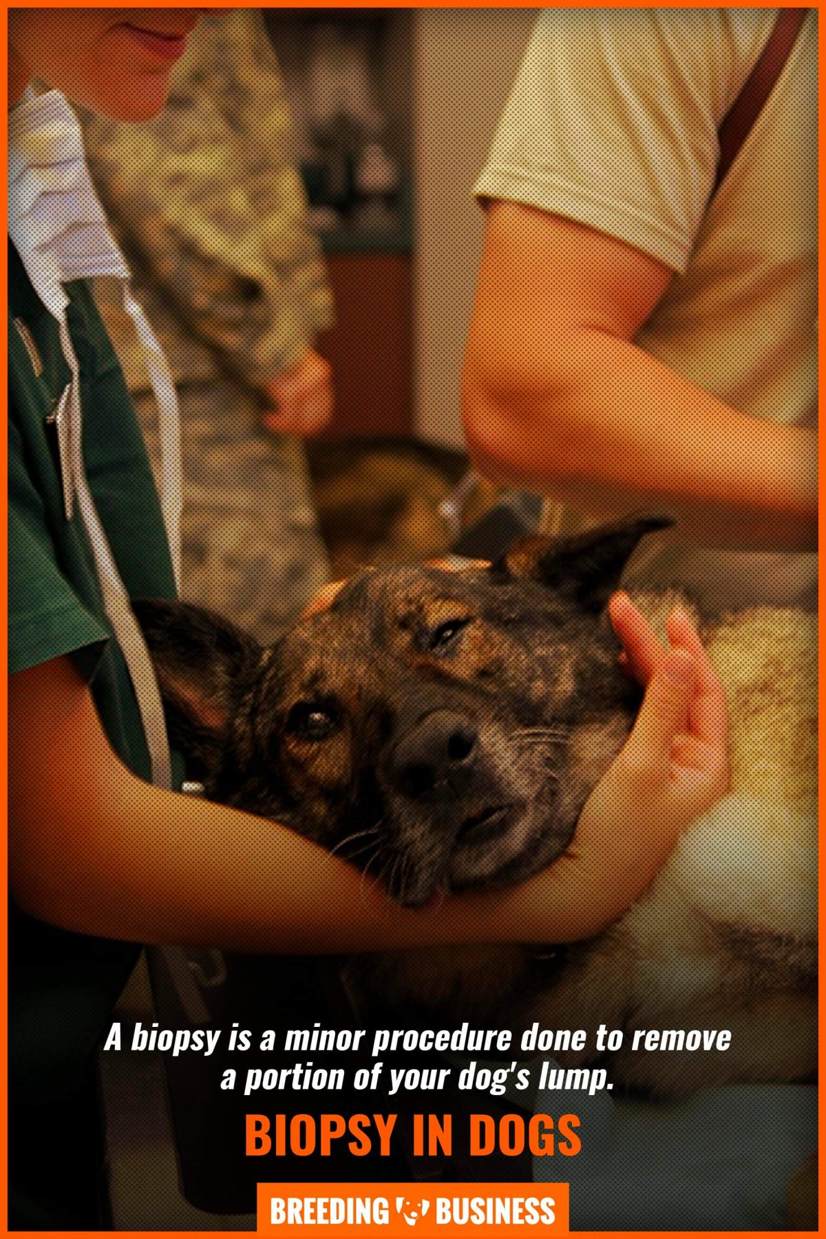
Biopsy
A biopsy is a minor procedure done to remove a portion of your dog’s lump. While fine-needle aspiration is usually enough to diagnose the cause of your pet’s lump, there are some cases where it’s not enough. If your dog undergoes a biopsy, they will receive sedation or general anesthesia. Small samples are sometimes taken using local anesthetic, and the whole mass may be removed rather than taking just a small sample. As with any medical decision, always discuss the pros and cons with your vet to choose the best treatment plan for your beloved pet.
Impression Smear
Impression smears are only taken from moist and ulcerated surface lesions. Using an impression smear, your vet can identify any inflammatory, neoplastic or other cellular infiltrates that might be inside the lump. If the lump isn’t contaminated, then any surface bacteria and yeasts can also be identified. While this technique has its uses, it can only sample surface exudates and not any deeper tissues. For this reason, an impression smear is better used alongside a biopsy to get an assessment of the lesion before processing the tissue sample. In this instance, the area around the excised lump sample is blotted to remove any blood that might contaminate the sample, and the exposed surface is applied to a clean slide.
Dog Lump Treatment
The type of treatment that your dog needs ultimately depends on the cause of their lump. If you notice that your dog has a lump or bump, be sure to check in with your vet for a diagnosis. Once a diagnosis is given, you may ask your vet about the different treatment options that would suit your dog.
If your dog’s lump is benign or early malignant, a lumpectomy may be performed. This is the surgical removal of the lump. Similarly, partial removal or cryosurgery can be done to reduce lumps that can’t be completely removed. For more aggressive malignant lumps, your vet may suggest radiation therapy or chemotherapy to shrink and kill cancer cells. Other types of therapy that might be used to treat your pet’s lump include laser therapy, immunotherapy, and multimodal therapy.
Dog Lumps – FAQ
Have any more questions about dog lumps? Feel free to consult our Frequently Asked Questions section for more details. As always, be sure to ask your vet for advice if you find a lump or bump on your dog.
Most cancerous lumps are typically hard or firm to the touch, and cannot be moved around under the skin. This is because they attach to the underlying tissues as they grow. However, it’s important to know that not all soft lumps are benign. Mast cell tumors (MCTs) resemble many other lumps and bumps and vary massively in appearance.
Some occur as small, moveable tumors under the skin. Others grow to become large and hairless. This highly variable presentation means that they may be mistaken for benign lipomas or warts, leading some owners to put off seeking veterinary advice.
The warning signs of a cancerous lump are very similar to those in humans. If your dog has a lump that grows rapidly, bleeds abnormally, feels hard, or causes itching, it’s possible that the lump is cancerous. Any changes in the lump’s size, color, shape, or ulceration are cause for concern. Because cancerous tumors can look and feel many different ways, it’s important to get your dog’s growth checked out by a vet as soon as possible, even if you suspect it to be benign.
Additional signs of cancer to look out for include wounds that don’t heal, swelling of a limb, abnormal bleeding, and unexpected weight loss. If you suspect that your dog might be sick, keep a close eye on their behavior. Different types of cancerous growths may cause different symptoms.
For example, limping and swelling in one leg can indicate bone cancer. As well as this, sudden abnormal behavior and seizures can indicate brain cancer. Melanoma can show itself through a swollen paw or a dark spot in the mouth.
Just like in people, there are many different types of lumps and bumps that can grow on your dog’s body! Your dog might develop hematomas, abscesses, granulomas, haemangiomas, hives, lipomas, sebaceous gland hyperplasia, histiocytomas, and carcinomas.
The list doesn’t stop there, extending to other growths such as skin tags, perianal adenoma, warts, and mast cell tumors. Because there are so many different growths in dogs, it’s important to seek veterinary advice if you find a new lump or bump.
While cysts and tumors can appear similar, there are several ways to tell them apart. Most importantly, a cyst is a sac filled with fluid, air, or tissue, a tumor is usually a solid mass of tissue. A cyst has a distinct membrane and is separate from any underlying tissues, whereas a benign or malignant tumor can attach itself to nearby tissues.
The causes of cysts and tumors are also different. A benign tumor can be caused by radiation exposure, genetic predisposition, and local trauma. Cysts, on the other hand, are typically the result of clogged sebaceous glands, infections, inflammatory conditions, and injuries that break the vessels.
There are many types of dog lumps that can afflict your pet, so it’s important to check in with your vet if you notice a new lump or bump. Whether the cause is a cyst or tumor, early detection of any underlying illnesses will give your pet the best prognosis possible.