
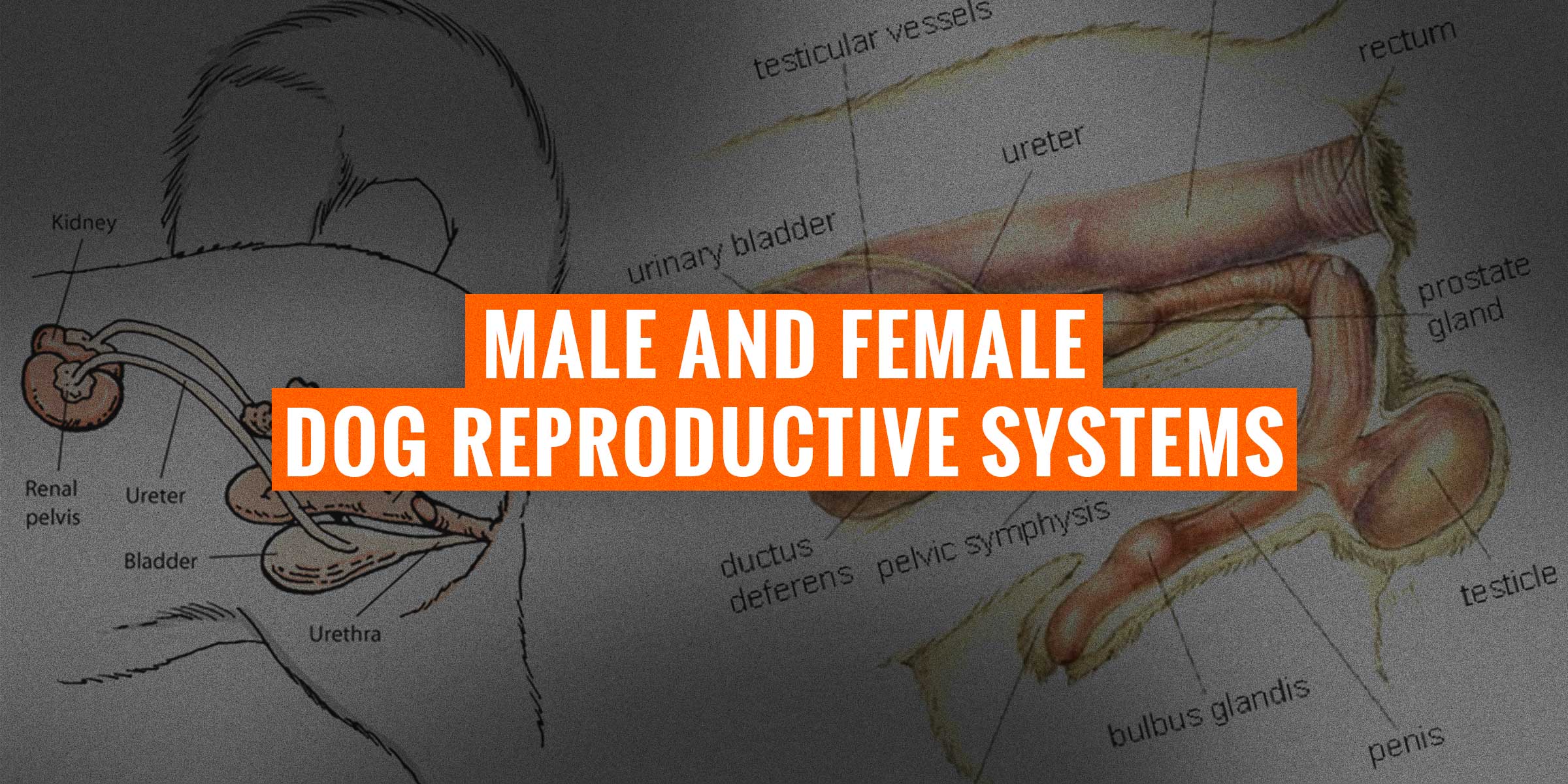
The dog reproductive system, also called canine genital system, is an interlinked system of sex organs within an organism which work together for the purpose of sexual reproduction. Such system includes obvious body parts such as the male’s penis and the female’s uterus, but also requires several hormones and other smaller but very functional organs.
Generally, the reproductive system is represented differently for female and male dogs since the organs differ very much. Here is a comprehensive guide covering:
- how both male and female reproductive systems work,
- what organs are they comprised of,
- which hormones govern them and to what extent, and
- improving your dog breeding program by understanding both reproductive systems.
This article is lengthy but we structured it in easily identifiable sections, one for each organ! Use our table of contents to jump from one section to the other.
Male Dog Reproductive System
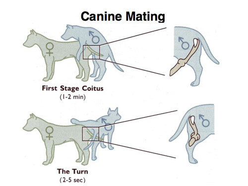
The canine reproductive system is, in essence, the system in which reproduction of the species is made through the act of coitus, or copulatory tie. The goal of the reproductive system is to produce adequate sperm and carry it into the female. The system consists of the dog’s genitalia and sexual organs that are solely responsible for preparing an organism for the act of mating with each organ maintaining its own function within the whole sexual system.
Sexual reproduction is accomplished through various periods in which both dogs, male and female, are prepared to mate. Much unlike the female dog who has distinctive heat cycles that distinguish her eagerness to mate, the male dog can be sexually aroused at any time during his lifetime and will usually oblige unless other physical factors keep him from doing so.
The dog reproductive system is made up of the reproductive anatomy. In the male, the main organs that are involved in sexual intercourse are the:
- penis,
- prostate gland,
- testicles,
- vas deferens or ductus, and
- sperm production.
When the male is aroused, these organs prepare for coitus by the arousal of the penis as the testicles are raised into position. During ejaculation, the sperm is transported via the vas deferens to the prostate gland where it is joined with essential, nourishing fluids then enter the urethra and finally to the penis. From the penis, the semen is directed into the uterus of the female for semination.
The penis is designed with two basic frameworks that work in unison for canine reproduction. The bulb-like structure at the base of the penis, called the glans penis, fills with blood and is used to hold the penis erect inside the vagina. The penis is simultaneously shielded and protected within the prepuce, while the inner bone helps to maintain the shape and position.
With that said, the male dog reproductive system is not exempt to outside threats, in fact, male dogs are susceptible to various reproductive disorders such as phimosis and cryptorchidism.
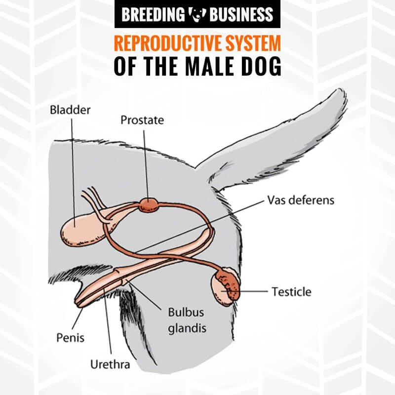
Bladder
The dog urinary bladder is a small sac located in the pelvic region above the pubic bone. The shape of the bladder resembles the shape of a pear when empty. It is here where urine is transported by the ureters from the kidneys. The bladder is what contains the urine in any given species. The urine is then transported into the urethra to the outside of the body in normal circumstances.
However, the urethra is also the same organ that transports the semen during ejaculation. In this case, when a male dog is sexually aroused, the urine is blocked, allowing for only semen to pass through and thus, allowing for the ejaculation to occur. A common bladder disorder in dogs, or simply in the lower urinary tract, can be caused by stones, spinal cord abnormalities, trauma or bladder inflammation.
Prostate
The male prostate gland functions mainly as a passageway for prostate fluid to flow. The fluid that flows via the prostate is for the nourishment and sheer protection of the semen. This fluid is literally squeezed through the urethra mixed in as it is expelled during the ejaculation phase of the sexual reproduction cycle. This course of action of expelling prostate gland fluid with semen is enabled by the male prostate gland muscle.
Without the prostate, semen cannot be properly and forcefully produced and excreted. The most prevalent canine prostate disorder in male dogs is Benign Prostatic Hyperplasia (BPH) which is marked by an increase of the weight and size of the gland with increasing age. This common disorder is often the cause of another common anomaly, an enlarged prostate, which can block the urethra by pressing down on it.
Penis
[pullquote-right]Inside the penis, small sponge-like tissue compartments allow blood to fill when the male is sexually aroused.[/pullquote-right]
The penis is the physical organ used in coitus and is the organ responsible for achieving the copulatory tie with the female. This organ is comprised of:
- the root,
- the base (shaft), and
- the cone-shaped end of the penis called the glans.
The root connects the penis to the abdomen and the glans is covered by a skin covering called the foreskin, which is often removed during circumcision. This glans is also where the tip of the penis is located to allow the expulsion of the semen. The penis of a male dog contains a V-shaped bone called the os penis (or baculum) that extends the penis’ length.
Inside the penis, small sponge-like tissue compartments allow blood to fill when the male is sexually aroused. This process causes the penis to stay erect during mating until the orgasm is reached, in which it becomes soft again. Unneutered male dogs who suffer from BPH are prone to bloody discharges of the penis.
Urethra
The urethra is the tube directly located inside the penis for easy passage of fluids. The fluids in a male’s anatomy need somewhere to easily pass through to be expelled, however, there needs to be an accommodating source that controls the flow of conflicting fluids, such as urine and semen. When the penis is erect, the urethra blocks the flow of urine to allow the easy passage of semen until ejaculation is complete. When the male is not aroused, urine is passed through.
There are many diseases that affect the urinary tract in male dogs including urethra, bladder, and prostate which are referred to as lower urinary tract diseases (LUTD). Urethral neoplasia is depicted by an overgrowth of mass cells inside or outside the urethra which is a common condition in male dogs and may require surgical intervention.
Bulbus Glandis
The bulbus glandis, also commonly referred to as the bulbourethral glands or the Cowper’s Glands are located on the sides of the urethra. The shape of these two structures are small, similar to the shape of a pea and are found just below the male prostate gland.
The main function of the bulbus glandis is to produce the fluids in the urethra that serve to neutralize the acidity of the environment that may develop from the residual fluids of urine inside the urethra. This gland also works to lubricate the urethra. The fluid is usually clear and very sleek allowing for easy slippage. Swelling of these glands are normal during arousal however some dogs may suffer from tumors.
Testicle
The testes, or gonads, are the small organs located inside the scrotum of the male sexual anatomy. Each testicle, or testes, is secured in place by the spermatic cord and come in two, however, some males have been known to contain just one which is usually rare. The two main functions of the testicles in the canine reproductive system are to produce the male sex hormone, testosterone, and the dog’s sperm.
Seminiferous tubules are coiled tubes inside the testes that assist in sperm cell production. Injuries to the testicles can cause testicular swelling, a common condition in male dogs, or cryptorchidism which is when the testes are not descended. Furthermore, the measurement and examination of the testicles can help one to determine any reproductive diseases and evaluate fertility.
Vas and Ductus deferens
The main function of the vas deferens, or also referred to as the ductus deferens, is to transport sperm from where it is stored into the urethra when the sperm is readily mature and ripe for semination.
The vas deferens is a long and thick tube, muscular in structure, that travels into the pelvic cavity from the epididymis. The ductus deferens comes in two, identical structures that function together in the same fashion. Epididymitis is a rare male sexual condition in dogs that is related to the abnormal operation of the vas deferens and coinciding organs.
Semen & Sperm
The semen is the viable liquid that is expelled from the urethra and penis during ejaculation. The semen is a protective fluid and is what carries the sperm, the reproductive cells.
The semen fluid contains spermatozoa, fructose, enzymes, and proteolytic and functions mainly as the carrying vehicle for the sperm cells. Common abnormalities and conditions in the male canine reproductive system related to semen are infertility issues where a male, for any reason, cannot impregnate a female. There is a vast array of various causes that can lead to infertility.
Hormones
The male reproductive system is comprised of hormones which act as the catalysts for cell activity. Reproductive cells and organs within the dog’s body are regulated by hormonal chemicals. There are three primary hormones that are involved in the male reproductive system:
- testosterone,
- luteinizing hormone (LH), and
- follicle-stimulating hormone (FSH).
Both LH and FSH are produced by the pituitary glands in the brain. The LH hormone stimulates testosterone production while the FSH hormone stimulates sperm production. Testosterone is responsible for producing the masculine qualities in the male including sexual drive, muscle and bone mass, and fat distribution. Some conditions can cause elevated sex hormones in males caused by factors such as uncastrated males or tumors, for example. Hormonal problems, whether it be a lack or overproduction, can lead to an array of symptoms such as hair loss, weight fluctuations, or increased thirst and urination. Always keep the hormones in check and regularly tested.
Female Dog Reproductive System
The canine reproductive system for females differs greatly from the male counterpart. Whereas a male may be stimulated at any time, a female must be in a designated heat period for copulation to flourish successfully. The female dog’s reproductive anatomy consists of:
- the ovaries (oviducts),
- the uterus,
- the cervix, and
- the vagina.
The production of unfertilized eggs is initiated inside the ovaries as they pass into the oviducts where they are fertilized by the male sperm. They are then transported to the uterus where the uterine body, and the left and right horns, are positioned for implantation of a fertilized egg to form into an embryo, firmly attached by the mother’s placenta.
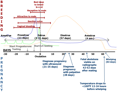
Heat Cycle
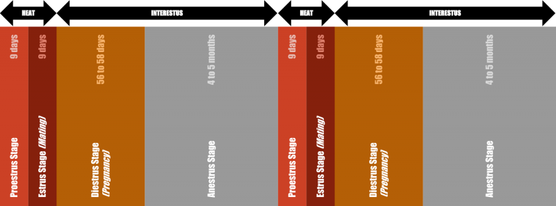
The reproductive hormones in a female’s body that are released by the ovaries and various structural glands help regulate the heat cycles of a female. A female dog will have her first heat cycle upon puberty between the age of 5 and 18 months, depending on the breed and size of the dog, and continue every 6 to 9 months of her life. The heat cycle is composed of 4 distinct phases and the timing may vary from one dog to the next:
- proestrus — the start of the heat cycle,
- estrus — the period the female is fertile,
- diestrus — the pregnancy, if successful, and
- anestrus — the rest or recovery from pregnancy.
Proestrus, the first stage, lasts 5 to 9 days and marks the beginning of the heat cycle. At this stage, the female may experience vaginal bleeding and the vagina well swell up. This stage also marks the time males begin to show interest, yet she is not engaging. The estrus stage lasts 5 to 9 days and is the prime time for a female to impregnate and she is receptive to a male’s advances where a copulatory tie is likely to occur. At this stage, the insemination of the sperm and egg will occur. The diestrus stage comes next and will last until the end of pregnancy where the embryo and placenta firmly attach to the uterus. Finally, the anestrus stage is marked by the in-between time between the cycles with no outward behavioral signs from the female.
We published a popular article about the various abnormal heat cycles that a female dog may a victim of.
Hormones
The female dog reproductive cycle is governed and constantly regulated by hormones in the body located both in the brain and the ovaries.
The hormones influence the fertility of the dog as well as regulate how the reproductive organs operate and function as well as their changes. In the female dog, the follicle-stimulating hormone (FSH) is located at the base of her brain in the pituitary gland and sends the signal for the development of the eggs in their follicles.
Eggs, once fully formed, will be able to form their own hormones. Estradiol 17B hormones are made by the follicles to dominate the follicular phase. Once matured, the luteinizing hormone (LH) is produced for the stimulation of the ovulation phase.
Progesterone and estrogen are the two main sexual hormones in a female dog and can be produced from the ovaries or the adrenal glands. Estrogen prepares the reproductive tract for breeding, assists in the motility of the female and male gametes, and affects her behavior to make her more attractive to males. Progesterone helps maintain a healthy pregnancy by providing adequate nutrition to the fetus and helps suppress the dam’s immune response to the “foreign” baby.
Ovaries
The ovaries of the female reproductive system, or female gonads, actually have two functions:
- production of the reproductive hormones progesterone and estrogen, and
- production of the eggs or oocytes.
The brain releases the Luteinizing Hormone due to the estrogen, which then causes the eggs to be released from the ovaries. The eggs inside the ovary are enclosed inside follicle cells which work to support the eggs. The ovaries in a female dog anatomy are situated close to the kidneys and are enclosed by the infundibulum tissue. It is in here where the ovaries are received for transport.
Ovaries are small, oval glands located on both sides of the uterus and are a major functional part of the system that transports ova to the fertilization site. When the female dog hits puberty, the form and size of the ovaries begin to change. The most common health issue related to the ovaries are ovarian cysts and tumors, or ovarian cancer. Ovarian tumors are commonly associated with non-spayed dogs.
Fallopian Tubes
The fallopian tubes are essentially where the fertilization and conception of an egg is established. Once an egg is fertilized in the fallopian tubes, it is then transported to the uterus for implantation. Because of this crucial function, this structure serves as an important facet of the canine reproductive system.
The uterine tubes, or oviducts, carry out their responsibilities in a matter of two days in a female dog and it is in these tubes that the egg begins its maturation phase. Many female dogs tend to suffer from common disorders that tend to show up as pathological changes known as lesions in the uterine/fallopian tube. Studies have shown that these lesions are generally characterized as disorders of growth, inflammation, cystic, and development. Problems that arise related to the fallopian tubes can eventually cause infertility in a female dog.
Uterus
After the fertilized egg has passed through the fallopian tubes it nestles into the uterus. Then, with the aid of the uterine wall where it firmly plants itself, the egg becomes ripe for development. However, in a female dog, as opposed to the human counterpart and other species, the uterus is made up of two horns that tend to be long, with a shorter body.
In addition to the uterus being the site for the implantation to occur, the female dog uterus also serves as the location in which the placenta and fetal development ensues. The uterus holds a pair of uterine horns that, together in unison, create the entirety of the uterus body. The uterus is then attached to the vagina where the opening of the cervix allows the passage of the fetus at the time of birth.
Young and middle-aged female dogs can suffer greatly from pyometra; however, this is more common in the older population. This condition is usually due to dogs undergoing many consecutive years of pregnancy-free oestrus heat cycles. Some dogs, after undergoing delivery, may experience subinvolution of the placental site, in which the normal process of the contractions of the uterus back to its normal size ceases to occur.
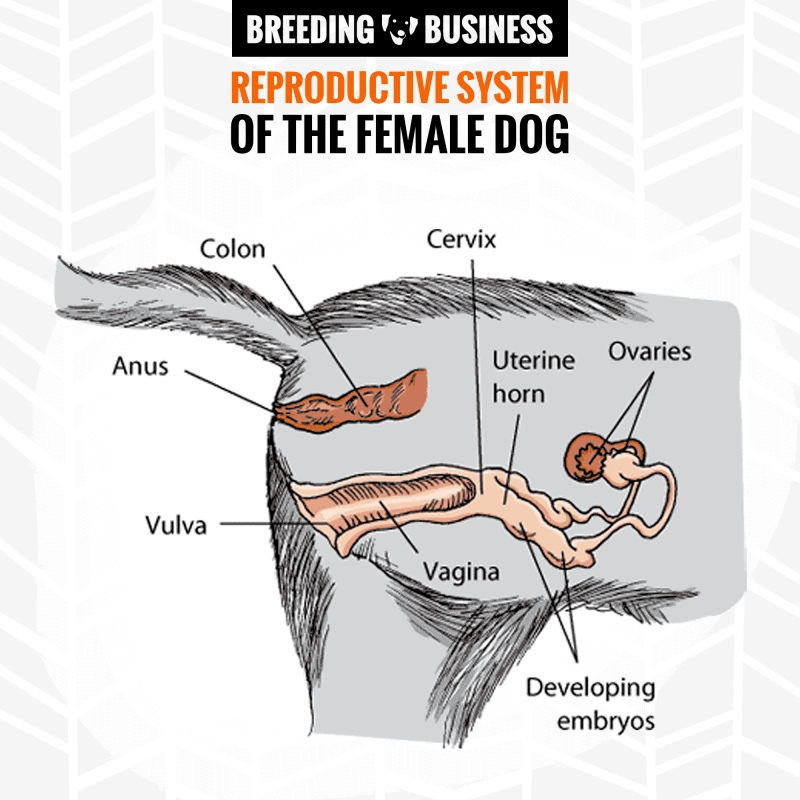
Cervix
The channel that serves as a basic midpoint between the uterus and the vaginal canal is the part of the female sexual reproductive system called the cervix. The cervix is a valve and serves as a junction between the uterus and the vaginal canal. It also serves the additional function of protecting the uterus from outside, foreign micro-organisms by closing up the birth canal during pregnancy. In addition, the cervix makes up part of the female genitalia.
A common physical ailment of the female dog related to the cervix is pyometra, which is normally associated with the uterus but also produces great effects on the cervix as well. Canine pyometra can be categorized as open cervix, or open pyometra, and closed cervix. Pyometra is a common condition in female dogs due to an accumulation of pus in the uterine lumen, usually showing up in a dog after a long bout of dominant progesterone.
Vulva
The dog’s vulva is the external opening to the female genitals. The vulva, like the cervix, makes up the area of the female sexual anatomy referred to as the genitalia. The vulva also consists of the sexual areas of the genitalia such as the clitoris, which is a sexual organ, and the double vertical lips outside the vagina. These two lips known as the outer lips, or the labia majora, and the small lips, or the labia menorah, function as a protective door.
In addition, the vulva also contains the opening to the urethra, therefore, assisting in the passage of urine. The most common sexual disorder in female dogs related to the vulva zone is vaginitis, or juvenile vaginitis, which is the inflammation of the vagina.
Mammary Glands

Mammary glands, or breasts, are designed for the sole purpose of milk production during pregnancy and milk delivery during the nursing of a litter of puppies. Unlike the genitalia which is specially designed for the pleasure and act of coitus in dogs, the mammary glands’ main responsibility is to provide nourishment to newborn puppies during the whelping phase of motherhood.
The mammary glands are located on the female dog’s underbelly and are lined up in two parallel rows consisting of 5 glands per line. Composed of glandular and connective tissue, each breast contains a nipple, or teat, for milk secretion through the mammary ducts. Usually, about 8 to 12 teats are common in a female dog.
Many lactating dogs suffer from a common condition called mastitis, which is an inflammation of the mammary gland caused by a bacterial infection. Agalactia, another known condition, is the failure of lactation in the mammary glands. Moreover, the most common tumors that female dogs suffer from are mammary gland tumors.